Research Topics
Effect of mechanical environment on collective cell migration using PDMS-based microfluidic device
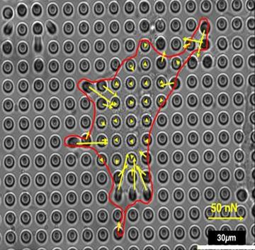
Collective cell migration is a high out-of-equilibrium process where cells are passively interacting with each other and exert an active force in response of external mechanical environment. These collective behavior plays a critical role in biological processes including morphogenesis, wound healing and cancer metastasis. In the past years, single cell migration has been extensively studies, however, the dynamic behavior of collective cell migration has not been well explained based on single cell movement. It is still under investigation how mechanical environment affects collective cell migration.
A substrate stiffness is one of the parameter to control the cell migration and traction forces at the cell-to-substrate interface. In this study, we present the effect of substrate rigidity on the collective cell movement using a PDMS-based microfluidic device. To provide difference in substrate stiffness, we fabricated arrays of oval-shaped micropillars incorporated in the device. Using time-lapse image sequence analysis, the influence of the micropillars substrate stiffness on collective cell migration rate was investigated. As a result, it was demonstrated that the cells in a group were able to sense and respond to alterations in the substrate stiffness.
Cell mechanotaxis using microchannel device and application to cancer cell migration
Over the last decade, we have seen a dramatic increase in the number of large, highly complex data sets being generated from biological experiments, quantifying molecular variables such as a cell, gene, protein, and metabolite abundance, microbiome composition, and population-wide genetic variation, to name just a few. Deep learning, an area of long-standing and growing interest in biological research, aims to address this complexity, providing next-level analyses that allow one to take new perspectives and generate novel hypotheses about living systems. Purpose of the study is deep learning in a new computation-based identifies for leader cell of cancer is decoded by Rac1. In this approach, identifying the leader cell of cancer-based on Immunofluorescent staining method, is an antibody, observing time-lapse imaging of Rac1. In the present study, Rac1 was originally identified GTPase which is responsible cell motility, and quantitatively identified the response functions that describe the conversion from the molecular activities to the morphological changes and mostly located in lamellipodium in the cell.
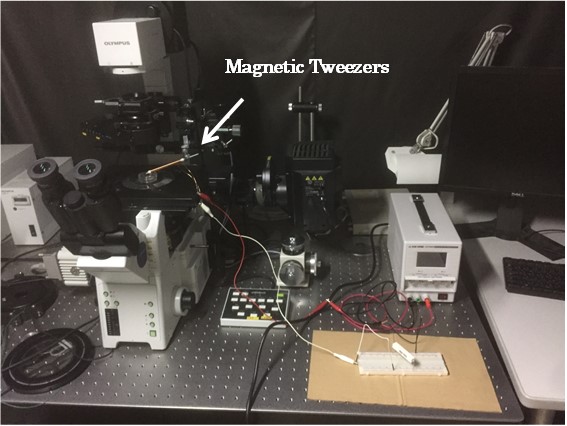
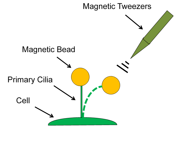
Investigation of the Function of Epithelial Primary Cilia as a Mechanosensor by Magnetic Tweezers-induced Mechanical Forces
In these decades, primary cilia have been getting attention for the function as a mechanosensor of mammalian cells. These organelles exist on the apical surface of cells one by one, and are believed to influence cell morphology, such as elongation and alignment when the cilia get external stimulation. Although numerous studies have been performed to estimate the actual function of primary cilia, little is known about their effect to cell remodeling. To investigate how primary cilia sense the external stimulation, I apply magnetic force onto the cilia with using magnetic trap method and observe the cell variation. A single magnetic bead is attached on the tip of fluorescent-stained primary cilia, and pulled by magnetic force generated from the point of magnetic tweezers. The behavior of primary cilia will be compared with that of previous studies.
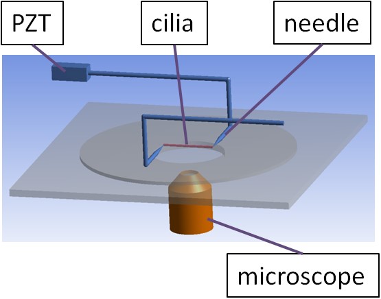
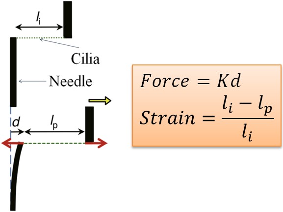
Measurement of mechanical property of primary cilia by stretching test
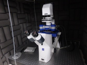
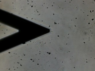
Primary cilia are hair-like organelles, which protrude from the surface of most cells. There are increasing interests on the properties of primary cilia. We try to measure mechanical property by stretching test using homemade stretching system (fig.1). Movement of piezo actuator is transferred to primary cilia via glass rod. Strain is defined as the ratio of total deformation to the initial length of primary cilia. Applied force on primary cilia is calculated by multiplying total deformation and spring constant of needle (fig.2). From relationship between strain and force, we aim to get mechanical property like young’s modulus.
Measuring Mechanical Property of Primary Cilia Using Atomic Force Microscope
Primary cilia are ubiquitous mammalian cellular substructures implicated in an ever-increasing number of regulatory pathways. Nonmotile primary cilia are slender subcellular structures that extend from the mother centrosome, are typically several microns long, and are used by eukaryotic cells to sense fluid flow. The widespread presence and conserved structure of primary cilia suggest they are a highly organized, functional organelle that plays a significant role in cell physiology and pathophysiology. However, our understanding of the functions of the primary cilia and its role in responding to environmental stimuli are still poorly understood. In this study, we isolate primary cilia from cultured Madin-Darby canine kidney cells(MDCK) and determine its mechanical properties using Atomic Force Microscope(AFM).
Development of Newly Designed Bioassay System for High-Throughput Single Cell Analysis
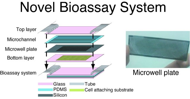
Over the last decade, microfluidics or “lab-on-a chip” technologies have been of great interests of a number of researchers as these technologies can be used as a biological assay system (e.g. cancer screening). Through an active collaboration with Dr Helen Andersson-Svahn at Royal Institute of Technology (Kungliga Tekniska hogskolan, KTH), we are developing a bioassay system consisting of an active microfluidic device integrated on the microwell plate for a high-throughput single cell analysis. This novel device can also be used for study of cell biomechanics under fluid shear stress.
Study on Measurement of Traction Forces Generated by Smooth Muscle Cells
It has been shown that intracellular structures rearrange according to changes in mechanical environments to which cells are subjected. In particular, cellular traction forces are believed to play an important role in the interactions between cells and their substrates, possibly involving the process of the cell remodeling. Although many studies have been performed to estimate cellular traction forces using microfabrication techniques, little is known of the effect of mechanical environments on the magnitude and direction of traction forces and hence how intracellular structures contribute to traction forces. This study addresses fundamental aspects of how these cytoskeletal structures contribute to smooth muscle cell mechanics.
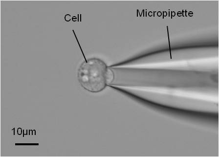
Mechanobiology of Chondrocyte Study of Chondrocyte in Response to Mechanical Stress
It is essential for cartilage health and application in cartilage tissue engineering to know chondrocyte mechanotransduction-the process by which cells sense and respond to mechanical signals. We study mechanical properties of chondrocytes by using a micropipette aspiration technique in which the surface of the cell is aspirated into a small glass tube. This technique can measure the Young’s modulus of cells and can be utilized to apply mechanical stress to cells. Chondrocytes can be isolated from bovine metacarpal-phalangeal joints by a process of sequential enzyme digestion. To investigate how they respond to mechanical stress, we measure the Young’s modulus of cells by applying suction pressure which is controlled by a syringe pump. The Young’s modulus was determined by a previous study.This project is collaborated with Dr. Martin Knight and Professor Dan Bader at Queen Mary, University of London (UK).

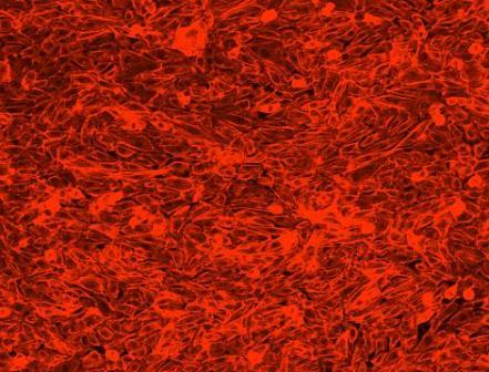
Mechanical responses of Myofibroblast to cyclic Stretching
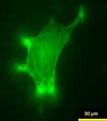
A wound healing is divided to three phases that are inflammatory phase, growth phase and mature phase. Myofibroblasts grow proliferously in the growth phase and align a certain direction. If they align a certain direction, a wound cures at early date, in contrast if they doesn’t align, a wound healing delays. But, it is unrevealed that what causes this alignment. we hypothesize that tensile stimulations of skins occurred by movements of dairy actions, and to reveal this hypothesis, we run a stretching experiment of myofibroblasts on the condition that a stretching rate is 1Hz and a stretching magnitude is 20%. After stretching, we observe an alignment of them with microscopy. At the same time, to observe their cytoskeleton, we run a fluorescence staining of actin filaments and an immunofluorescence staining of vinculins.
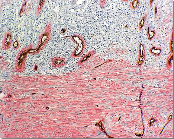
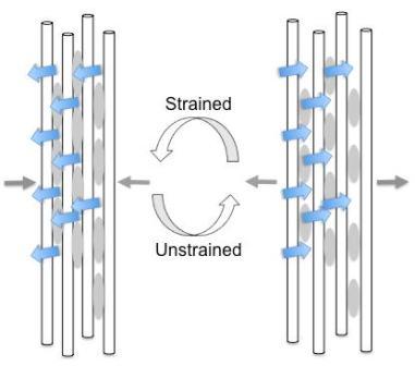
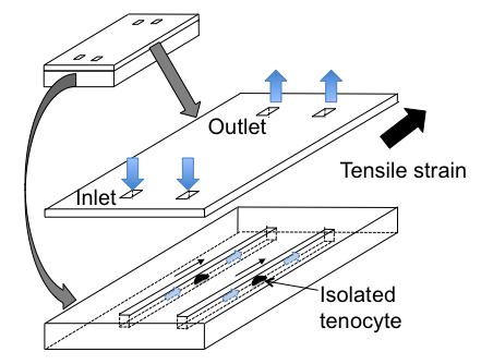
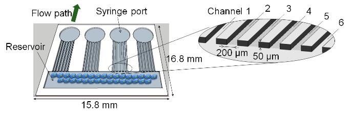
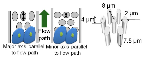
Mechanobiological Responses of Tenocytes to Interstitial Fluid Flow with/without the Presence of Cyclic Tensile Strain
Tendon is collagenous, soft connective tissue in articular joint, predominantly subjected to tensile loading. Tendon possesses water for approximately 60% of its wet weight. This water imposes fluid shear stress to tendon cells, tenocytes, when tendon matrix is stretched. Thus, local mechanical environment of tenocyte includes both tensile strain and fluid shear stress. In conjunction with MEMS techniques, this project develops a microfluidic device which enable to apply controlled tensile strain and fluid shear stress to isolated tenocytes in vitro, and examine their synergetic effects on tenocyte mechanotransduction events and associated mechano-regulation of tenocyte metabolism. This research project is in collaboration with Prof. James Wang at University of Pittsburgh.